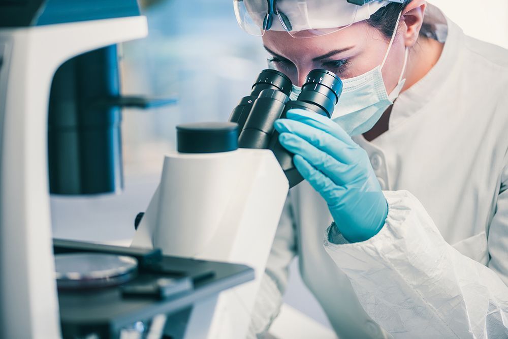
Direct Microscopic Examination and Evaluation in Superficial Fungal Infections
Microscopic examination is a simple, fast and inexpensive method used to diagnose superficial fungal infections (dermatophytes, candida infections and pityriasis versicolor).
Application
1) The patient should be informed about the procedure to be performed.
2) Gloves can be used for protection while working around the oral mucosa and genital area during the procedure.
3) Sampling can be made from skin, hair, nails, oral and genital mucosa.
When sampling the skin, dandruff is collected by scraping off the blunt-handled scalpel, a scalpel tip, or preferably the edge of a slide onto another slide.
In nail involvement, sampling is done with dandruff under the nail.
In case of scalp or beard involvement, a few hair samples that appear broken and structured on the lesion, as well as the squamae in the lesion area, are pulled with a forceps to be examined. The easy removal of the hair pulled with the inflammatory type forceps indicates that it is a suitable specimen for examination. In the beard clipper, the hair and crustal structures in the lesion area can be examined.
4) The material collected on the slide is brought together and covered with a coverslip.
5) On the material collected on the slide or after the coverslip is covered, 10-30% KOH is dripped from its edge with a dropper.
6) The prepared sample is left on a paper towel for 15-30 minutes, either open on a paper towel or in a petri dish with a wet sponge, so that the KOH dissolves the keratin and the fungal structures made of cellulose become visible.
7) The presence of fungus is determined by not examining the sample under a microscope.
In cases where a definitive diagnosis cannot be made by direct microscopic examination and the fungus genus needs to be determined, and in researches, culture method can be applied.
